Picture of Health
Today, several types of imaging technology help doctors see inside your body to diagnose disease. Here’s a closer look at a few of these.
One day in 1895, a German physicist named Wilhelm Röntgen was doing experiments with electron beams in a gas discharge tube. While projecting the beams onto a fluorescent screen, he saw a silhouette of the bones of his hand. By accident, he had discovered X-rays—and opened the modern era of medical imaging.
Today, several types of imaging technology help doctors see inside your body to diagnose disease. Here’s a closer look at a few of these:
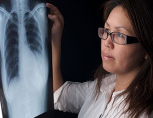
- X-RAYS are beams of electromagnetic energy like visible light rays, but with a different wavelength. They generate a film image in which hard tissue shows up better than soft because it’s made up of larger atoms. A mammogram is a low-dose X-ray exam of the breasts that is used to spot tissue irregularities that may be tumors.
- DEXASCAN, or dual-energy X-ray absorptiometry, combines two beams of very low-dose X-rays to measure bone density and help assess the risk of osteoporosis.
- COMPUTED TOMOGRAPHY (CT) scans use a number of X-ray images to assemble a three-dimensional picture of your internal organs. Lying on a platform, you move slowly through a machine that looks like a huge donut tipped on its side. An X-ray tube is mounted on a revolving ring inside the “donut,” with X-ray detectors facing it. In a spiral fashion the machine collects a series of “slices,” which the computer puts together.
Saint Peter’s University Hospital offers a 64-slice CT system that minimizes the amount of radiation that is needed, says Steven Schonfeld, M.D., of University Radiology Group and chairman of the hospital’s Department of Radiology. “It lets us do very high-quality studies through- out the body with comparatively smaller radiation doses.”
CT scans help diagnose cancer, among other conditions. And the new machines can be used for CT angiography, a less invasive alternative to catheter angiograms used in evaluating heart patients.
- ULTRASOUND is used in obstetrics, cardiology and urology. Similar to a submarine’s sonar, it’s based on the principle of echolocation, which whales and dolphins use to navigate. As a technician moves a tool called a transducer over the body, high-frequency sound waves are emitted. They bounce off the parts to be imaged and back to the transducer, which sends data into a central computer to create a moving picture visible on a monitor. Today, some ultrasound equipment can be used to create three- dimensional images.
- MAGNETIC RESONANCE IMAGING (MRI) uses powerful magnets to create an interior picture that helps doctors diagnose conditions from tumors to tendinitis. It too takes “slice” images, but unlike conventional CTs it can take them of body parts in any direction, not just horizontally. An MRI machine directs a radiofrequency pulse at the tissue to be imaged. This pulse causes protons in hydrogen atoms within the tissue to move in response to changes in magnetic fields, and normal and abnormal tis- sue react differently. When the protons move, they release excess energy the system’s computer uses to create the image.
MRIs highlight soft body tissues. A breast MRI, for example, captures multiple images of breast tissue. It may be done in circumstances—especially with dense breast tissue, for example—when there’s a need for more information than an ultrasound or mammogram can provide.
At Saint Peter’s, says Dr. Schonfeld, a large percent- age of scans are evaluated by fellowship-trained radiologists who specialize in a particular area, such as pediatric radiology or neuroradiology, and therefore bring more specific experience to bear.
“There’s a radiology resident and an attending radiologist available to review cases 24/7,” he says.